


Una nota sobre estos materiales de archivo
Esta página conserva apuntes de estudio elaborados durante cursos anteriores. No se ha revisado exhaustivamente para reflejar los estándares clínicos, científicos, de evaluación o normativos actuales. Se proporciona únicamente con fines educativos e históricos, no como consejo médico, guía de tratamiento ni sustituto de los materiales del curso ni de las directrices sanitarias oficiales.
Cerebro y nervios craneales
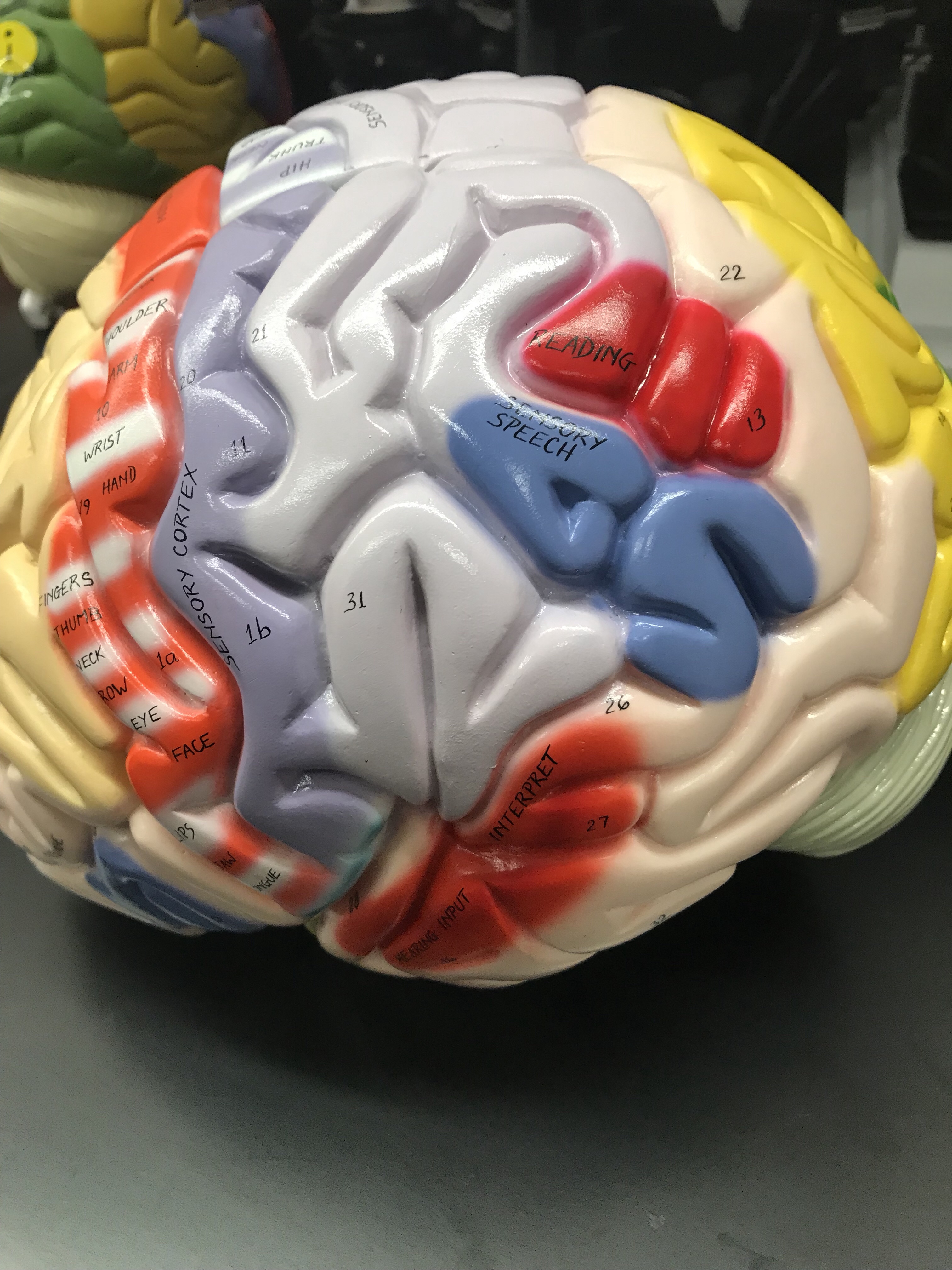
El sistema nervioso consta del Sistema Nervioso Central (SNC) y el Sistema Nervioso Periférico (SNP). Aferente: Sensorial, transporta impulsos hacia el SNC. Conduce hacia el centro. Lleva hacia adentro a un órgano central (por ejemplo, el cerebro) o sección, como los nervios que conducen impulsos desde la periferia del cuerpo al cerebro o la médula espinal. Son sensoriales. Eferente: Motor, transporta impulsos desde el SNC. Conduce o progresa desde el SNC o un sitio específico. Impulso nervioso hacia afuera del SNP. El Sistema Nervioso Central está compuesto por:- Cerebro
- Médula espinal
Combina información de todo el cuerpo y coordina la actividad en todo el organismo. Utiliza 201 TP3T del oxígeno que respiramos. Se estima que hay 100 mil millones de neuronas, cada una conectada a muchas más. Hay cuatro lóbulos principales: parietal, temporal, occipital y frontal. El cerebro está protegido por el cráneo. La médula espinal recorre la columna vertebral. El cerebro está alojado dentro de las meninges. La médula espinal transporta información entre el cuerpo y el cerebro. Se conecta al SNP (piel, músculos y articulaciones).
El lóbulo temporal es importante para procesar la información sensorial y asignarle un significado emocional: recuerdos a largo plazo; lenguaje (algunos).
El lóbulo occipital se encarga del procesamiento visual.
El lóbulo parietal integra información sensorial, incluyendo el tacto, la conciencia espacial y la navegación; procesamiento del lenguaje (en cierta medida).
El lóbulo frontal contiene la mayor parte de la dopamina; neuronas sensibles; involucradas en la atención; recompensa, memoria a corto plazo, motivación y planificación.
El cerebro es el módulo de control central del cuerpo. Es el órgano más complejo. Algunas secciones están especializadas. Otras funciones implican el trabajo en conjunto. Hay doce nervios. Ocho de ellos parecen estar a lo largo del tronco encefálico. Las células de la neuroglia dan soporte al tejido nervioso. El sistema nervioso periférico está compuesto por:- Todos los nervios y ganglios fuera del cerebro y la médula espinal.
 El tronco encefálico tiene tres partes: mesencéfalo, protuberancia anular y bulbo raquídeo. Cada una de ellas está compuesta por varias partes que tienen funciones específicas. Mesencéfalo
- El pedúnculo cerebral son axones que transfieren información
- Corpora quadrigemini 4 unidades
- El colículo superior se encuentra en la parte posterior del mesencéfalo. Recibe señales visuales de la retina de los ojos; ayuda con la orientación de los ojos y la cabeza; reflejos visuales.
- Los colículos inferiores se localizan posteriormente al mesencéfalo. La capa inferior procesa múltiples señales de diversas partes del cerebro. Su función principal es la integración de señales, el reconocimiento de frecuencias y la discriminación de parches; reflejos auditivos.
- Vías o tractos neuronales; centros respiratorios que ayudan al bulbo raquídeo; transmite información al diencéfalo.
- Centros cardíaco, respiratorio, del vómito y vasomotor (regulación de la presión arterial); es una zona vital; se extiende por debajo del cráneo. Interviene en funciones involuntarias como el vómito, la respiración, el estornudo y el mantenimiento de la presión arterial correcta.


Médula espinal y nervios espinales
Existen treinta y un nervios espinales. Estos se componen de los engrosamientos cervical y lumbar, el cono medular, la cola de caballo y el filum terminal. El engrosamiento cervical se extiende desde C4 hasta T1 y se dirige a las extremidades superiores a través de los plexos braquiales. El engrosamiento lumbar (también llamado engrosamiento lumbosacro) se ubica desde T9 hasta T12. Forma los plexos sacro y lumbar, que se dirigen a las extremidades inferiores. El cono medular, que se encuentra en el extremo de la médula espinal, tiene forma de cono. Le sigue un haz de nervios llamado cola de caballo antes de terminar en el filum terminal. El nervio frénico inerva la médula espinal y ayuda al diafragma. Se encuentra dentro del plexo cervical de la columna vertebral, entre C3 y C5. Recuerda el dicho: "Los nervios cervicales 3, 4 y 5 mantienen vivo el diafragma".plexo raquial Contiene cinco nervios principales: el nervio axilar (región del hombro), el nervio musculocutáneo (parte anterior del brazo), el nervio mediano (parte más anterior del antebrazo), el nervio cubital (la mayoría de los músculos de la mano) y el nervio radial (todos los músculos posteriores del brazo y el antebrazo). El plexo braquial se encuentra entre C5 y T1. Los nervios torácicos (o intercostales) no tienen plexos. Sin embargo, el plexo lumbar tiene dos nervios que inerva: el nervio femoral, que va al cuádriceps, y el nervio obturador, que va a los músculos aductores (parte medial del muslo). El plexo sacro contiene el nervio más grande del cuerpo, llamado nervio ciático, que va a los isquiotibiales (parte posterior del muslo), y el nervio pudendo, que va a los músculos del perineo de la zona genital. La médula espinal está compuesta por la raíz dorsal y ventral, así como por la médula espinal. La raíz dorsal consta de axones sensitivos somáticos y autónomos que entran en la médula espinal a través de ella. La protuberancia en el lateral de la raíz se llama ganglio de la raíz dorsal. Está compuesto por cuerpos celulares de neuronas sensitivas. La raíz ventral también tiene axones somáticos y autónomos, pero son sensitivos. Salen de la médula espinal a través de la raíz ventral. El nervio espinal es donde convergen las raíces ventral y dorsal y sale por el foramen intervertebral. Se llama mixto porque contiene fibras sensitivas y motoras. La rama de la raíz dorsal inerva la piel y los músculos profundos de la espalda y el tronco. La rama de la raíz anterior inerva los músculos y la piel de los brazos y las piernas, así como las partes anteriores y laterales del cuerpo. Las ramas meníngeas trabajan con las vértebras y sus ligamentos, así como con las meninges y los vasos sanguíneos de la médula espinal. La médula espinal tiene el surco mediano posterior y la fisura mediana anterior, así como el canal central y las comisuras gris y blanca. La fisura es un surco ancho, mientras que el surco es estrecho. El canal central se extiende a lo largo de la médula espinal. La comisura gris y blanca une los lados izquierdo y derecho. Las columnas de sustancia blanca (o funículos) contienen el tracto sensitivo ascendente y el tracto motor descendente, que consta de haces de axones del SNC. Hay un lado anterior, lateral y dorsal de las columnas. La sustancia gris tiene astas ventral, dorsal y lateral. Los cuerpos celulares de las motoneuronas somáticas se encuentran en el asta ventral. El asta dorsal contiene los cuerpos celulares de las interneuronas. En el asta lateral del SNA se encuentran los cuerpos celulares de las motoneuronas, excepto en la región cervical.


 Existen tres tejidos conectivos protectores que recubren cada médula espinal:
- Epineuro que rodea todo el nervio
- Perineuro que rodea el fascículo (que es un haz nervioso más pequeño)
 NOTA: LOS NERVIOS SE REPRESENTAN GENERALMENTE EN NÚMEROS ROMANOS.
CN#1 El nervio olfativo es para el olfato.
CN#2 El nervio óptico es para la vista (bastones y conos) - visión
CN#3 El nervio oculomotor mueve los párpados y los globos oculares CN#4 El nervio troclear mueve los globos oculares CN#5 El nervio trigémino para los músculos faciales (por ejemplo, masticar) CN#6 El nervio abducens mueve los globos oculares CN#7 El nervio facial para el gusto, las lágrimas, la saliva y la expresión facial CN#8 El nervio vestibulococlear o acústico es para la audición (nervio vestibular: equilibrio; nervio coclear: audición) CN#9 El nervio glosofaríngeo es un nervio sensoriomotor: deglución, saliva, gusto, vísceras CN#10 El nervio vago controla el SNP: control motor del corazón y las vísceras, sensación del tórax, la faringe y las vísceras abdominales CN#11 El nervio accesorio mueve la cabeza y los hombros CN#12 El nervio hipogloso trabaja los músculos de la lengua (habla y deglución), Algunos músculos esqueléticos, algunas vísceras, sensibilidad de la piel y las vísceras. Hay ocho pares de nervios espinales cervicales, doce pares de nervios espinales torácicos, cinco pares de nervios espinales lumbares, cinco pares de nervios espinales sacros y un par de nervios espinales coccígeos.
NOTA: LOS NERVIOS SE REPRESENTAN GENERALMENTE EN NÚMEROS ROMANOS.
CN#1 El nervio olfativo es para el olfato.
CN#2 El nervio óptico es para la vista (bastones y conos) - visión
CN#3 El nervio oculomotor mueve los párpados y los globos oculares CN#4 El nervio troclear mueve los globos oculares CN#5 El nervio trigémino para los músculos faciales (por ejemplo, masticar) CN#6 El nervio abducens mueve los globos oculares CN#7 El nervio facial para el gusto, las lágrimas, la saliva y la expresión facial CN#8 El nervio vestibulococlear o acústico es para la audición (nervio vestibular: equilibrio; nervio coclear: audición) CN#9 El nervio glosofaríngeo es un nervio sensoriomotor: deglución, saliva, gusto, vísceras CN#10 El nervio vago controla el SNP: control motor del corazón y las vísceras, sensación del tórax, la faringe y las vísceras abdominales CN#11 El nervio accesorio mueve la cabeza y los hombros CN#12 El nervio hipogloso trabaja los músculos de la lengua (habla y deglución), Algunos músculos esqueléticos, algunas vísceras, sensibilidad de la piel y las vísceras. Hay ocho pares de nervios espinales cervicales, doce pares de nervios espinales torácicos, cinco pares de nervios espinales lumbares, cinco pares de nervios espinales sacros y un par de nervios espinales coccígeos.
Hormonas
 Encontré esta canción genial que te ayudará a aprender sobre el sistema endocrino. MCAT musical: Hormones Blues
| Hipotálamo | Hormonas liberadoras/inhibidoras, TRH, GnRH, CRH, PIH, etc. | Regula la glándula pituitaria |
| Hipófisis anterior | hGH | Síntesis de IGF, promueve el crecimiento. |
| Hipófisis anterior | TSH | Síntesis y secreción de la hormona tiroidea |
| Hipófisis anterior | FSH | Secreción de estrógenos, producción de espermatozoides y ovocitos |
| Hipófisis anterior | LH | Ovulación; secreción de estrógeno, progesterona y testosterona. |
| Hipófisis anterior | MSH | Secreción y producción de leche |
| Hipófisis anterior | ACTH | Estimula la secreción de glucocorticoides (cortisol) de la corteza suprarrenal. |
| Hipófisis anterior | MSH | pigmentación de la piel |
| Hipófisis posterior | ADH | Aumenta la reabsorción de agua; disminuye la formación de orina; aumenta la presión arterial. |
| Hipófisis posterior | Antiguo Testamento | Estimula las contracciones uterinas y la bajada de la leche. |
| Tiroides | Tetralodotironina (T4) | Aumenta el metabolismo, la temperatura corporal y la producción de ATP. |
| Tiroides | Trilodotironina (T3) | Aumenta el metabolismo, la temperatura corporal y la producción de ATP. |
| Tiroides | Calcitonina | Disminuir los niveles de calcio en sangre |
| Paratiroides | PTH | Aumentar los niveles de calcio en sangre |
| corteza suprarrenal | Aldosterona | Niveles de Na y K; regula la presión arterial y el volumen sanguíneo. |
| corteza suprarrenal | Cortisol | Aumenta los niveles de glucosa, tiene efectos antiinflamatorios, aumenta la resistencia al estrés y deprime el sistema inmunitario. |
| Corteza suprarrenal | Andrógeno (DHEA) | Cambios en la pubertad; conversión a estrógeno y estimulación de la libido en las mujeres. |
| Médula suprarrenal | Epinefrina | Respuesta al estrés, promueve la respuesta de lucha o huida. |
| Médula suprarrenal | Norepinefrina | Respuesta al estrés, promueve la respuesta de lucha o huida. |
| Páncreas: Células alfa | Glucagón | Aumento de los niveles de glucosa en sangre |
| Páncreas: Células beta | Insulina | Disminuir los niveles de glucosa en sangre |
| ovarios | Estrógeno | Regula el ciclo menstrual, estimula el desarrollo de las características sexuales femeninas. |
| ovarios | Progesterona | Regula el ciclo menstrual, estimula el desarrollo de las características sexuales femeninas. |
| Testículos | Testosterona | Producción de esperma, características sexuales masculinas |
| Glándula pineal | Melatonina | Reloj biológico |
| Timo | Timosina | Maduración de los linfocitos T (glóbulos blancos) |
- Zona glomerular que secreta mineralocorticoides, entre los que encontramos la aldosterona.
- La zona fasciculada es la más grande. Produce glucocorticoides (cortisol). Un exceso puede causar el síndrome de Cushing, y una deficiencia, la enfermedad de Addison.
- La zona reticular produce gonadocorticoides (andrógenos), que aumentan el deseo sexual en las mujeres después de la menopausia.
Sistema digestivo
Vídeo musical sobre el sistema digestivo por video musical biológico El sistema digestivo se extiende desde la boca hasta el ano. La boca se utiliza para la masticación, que es el proceso de triturar los alimentos. Está compuesta por epitelio escamoso estratificado multicapa. Hay tres pares de glándulas salivales que secretan saliva, la cual contiene bicarbonato. Mantiene un pH entre 6,5 y 7,5. Mediante la peristalsis, el bolo alimenticio (una bola de alimento masticado) pasa de la boca al estómago. La peristalsis es el movimiento de los alimentos a través del tracto digestivo mediante contracciones. Los alimentos generalmente permanecen en el tracto digestivo durante tres días. La glándula parótida se encuentra delante y debajo de la oreja. Secreta una sustancia acuosa y transparente llamada amilasa, que descompone los carbohidratos. El conducto de Stensen está conectado a ella. La segunda glándula se llama submandibular y se encuentra en la superficie interna de la mandíbula. Secreta una sustancia serosa y algo de moco, y el conducto de Whatman está conectado a ella. Las células del tracto digestivo se renuevan cada tres días. La mayoría de estas células son epitelio columnar simple. El tracto digestivo consta de cuatro capas.
Cuatro capas del tracto digestivo
- La mucosa es la capa más interna del tracto digestivo. Está compuesta principalmente por epitelio y células mucosas. La luz intestinal es el conducto central del tracto digestivo.
- La submucosa es la segunda capa. Contiene vasos sanguíneos, nervios y vasos linfáticos. Los vasos linfáticos son los vasos linfáticos.
- A continuación, vienen dos capas de músculo.
- La capa más interna se llama fibras circulares
- Fibras longitudinales
- La serosa es la capa más externa.
- Peritoneo visceral que recubre los órganos
- Peritoneo parietal que recubre la cavidad peritoneal
- Evita el sonido nasal excesivo.
- Mantiene la garganta lubricada.
- Ayuda a evitar que la comida suba por la nariz y facilita el drenaje de la mucosidad.
- Reflejo nauseoso
- Ayuda en las respuestas inmunitarias
 El estómago está formado por tres capas musculares: oblicua, circular y longitudinal. La oblicua es la capa más interna. El estómago está revestido de células epiteliales columnares. En su interior, se encuentran los pliegues gástricos, también formados por células epiteliales columnares. Básicamente, los pliegues gástricos son surcos que facilitan la digestión. El agua, la glucosa, las sales, el alcohol y los fármacos son absorbidos por el estómago (absorción gástrica).
El estómago contiene tres glándulas gástricas diferentes que comienzan en el inicio del estómago y terminan en la luz del estómago. secreta jugo gástrico y moco.Cada glándula tiene una función específica. La glándula cardíaca, ubicada en la parte superior, y la glándula pilórica, ubicada en la parte inferior, secretan moco. Las glándulas gástricas intermedias son las que realizan la mayor parte de la secreción. Están compuestas por tres tipos de células: principales, parietales y del cuello mucoso. Las glándulas gástricas intermedias secretan HCl o ácido clorhídrico, necesario para activar ciertas enzimas. Dra. Iram Tassaduq Tiene una bonita presentación de diapositivas computarizada sobre el estómago. Las diversas secreciones de las glándulas gástricas son:
El estómago está formado por tres capas musculares: oblicua, circular y longitudinal. La oblicua es la capa más interna. El estómago está revestido de células epiteliales columnares. En su interior, se encuentran los pliegues gástricos, también formados por células epiteliales columnares. Básicamente, los pliegues gástricos son surcos que facilitan la digestión. El agua, la glucosa, las sales, el alcohol y los fármacos son absorbidos por el estómago (absorción gástrica).
El estómago contiene tres glándulas gástricas diferentes que comienzan en el inicio del estómago y terminan en la luz del estómago. secreta jugo gástrico y moco.Cada glándula tiene una función específica. La glándula cardíaca, ubicada en la parte superior, y la glándula pilórica, ubicada en la parte inferior, secretan moco. Las glándulas gástricas intermedias son las que realizan la mayor parte de la secreción. Están compuestas por tres tipos de células: principales, parietales y del cuello mucoso. Las glándulas gástricas intermedias secretan HCl o ácido clorhídrico, necesario para activar ciertas enzimas. Dra. Iram Tassaduq Tiene una bonita presentación de diapositivas computarizada sobre el estómago. Las diversas secreciones de las glándulas gástricas son:
- Pepsinógeno: secretado por las células principales y forma pepsina.
- La pepsina se forma cuando el HCl reacciona con el pepsinógeno. Ayuda en la digestión.
- El ácido clorhídrico (HCl) es secretado por las células parietales. Reacciona con el pepsinógeno.
- Moco: secretado tanto por las células caliciformes como por las glándulas mucosas. El moco se utiliza para proteger la mucosa. Es alcalino.
- Factor intrínseco: secretado por las células parietales, ayuda en la absorción de la vitamina B12, necesaria para la producción de sangre. La anemia perniciosa es causada por la falta de vitamina B12.
 El ciego es la bolsa ciega que se encuentra en la unión del intestino grueso y el intestino delgado. Se considera la primera parte del intestino grueso. El apéndice/vermiforme se encuentra en la parte inferior derecha del abdomen. La válvula ileocecal es una válvula muscular esfinteriana ubicada entre el íleon y el ciego. Aproximadamente dos litros de líquido entran a la válvula a través de la válvula ileocecal. Las tenias del colon son tres bandas musculares que se encuentran en la pared del colon. Las haustras son bolsas que se forman como resultado de las tenias del colon. Se proporcionará información sobre esto al final. Los apéndices epiploicos son pequeñas bolsas en el peritoneo que están llenas de grasa. Dentro del colon, hay numerosas células mucosas. Se pueden encontrar bacterias en el colon. Descomponen la celulosa para obtener energía (un carbohidrato) y sintetizan vitamina K, B12, tiamina y riboflavina. La colostomía es un procedimiento quirúrgico en el que se crea un orificio para sacar el intestino grueso del cuerpo para que el estoma pueda drenar en una bolsa. La diarrea (o espasmo peristáltico) es la condición en la que se tienen tres o más deposiciones al día. Generalmente dura unos pocos días y desaparece tan rápido como aparece. La disentería es una infección intestinal que causa la expulsión de heces con sangre. Puede ser causada por un parásito o una bacteria. Existen cuatro tipos de simbiosis: Mutualismo, comensalismo, parasitismo y amensalismo.El comensalismo se denomina camarilla. Se da cuando un organismo recibe beneficios de otro sin afectarlo de ninguna manera. El mutualismo se da cuando ambos se benefician de su asociación. El parasitismo se da cuando el parásito daña al huésped. El amensalismo es muy raro. Ocurre cuando un organismo no recibe ningún beneficio directo de la relación. Taenia saginata es una tenia bovina, mientras que Taenia solium es una tenia porcina. Además, Ascaris lumbricoides es un nematodo. Diphyllobothrium latum es un mamífero que se alimenta de peces. La tenia del pez puede alcanzar hasta 18 metros de longitud. Es un parásito intestinal que absorbe la vitamina B12 de su huésped. Entamoeba histolytica es un parásito intestinal que causa la disentería amebiana. Entamoeba gingivalis es un comensal de las encías. Vive en la boca de una persona y se reproduce asexualmente. Giardia intestinalis es un parásito del intestino delgado que causa la giadiasis. La cirrosis es causada por hígado graso y/o consumo excesivo de alcohol. Las hepatitis A, B, C y D afectan al hígado. Las hepatitis A y B se transmiten principalmente por alimentos o utensilios contaminados. Las hepatitis C y D pueden transmitirse por el semen.
Sangre
El volumen sanguíneo promedio de un ser humano es de aproximadamente 5 litros. La sangre está compuesta por células, proteínas, oxígeno y nutrientes, además de otros componentes. El hematocrito se compone de plasma, eritrocitos, leucocitos y trombocitos. El plasma constituye el 551% del volumen sanguíneo total. Representa la mitad del volumen sanguíneo. Está compuesto en un 90% de agua, mientras que el 101% restante son proteínas, minerales, productos de desecho, factores de coagulación, hormonas e inmunoglobulinas. Se separa del resto de la sangre mediante centrifugación. Los eritrocitos (glóbulos rojos) se almacenan en el bazo. Son discos bicóncavos que carecen de núcleo y viven aproximadamente 120 días. Transportan oxígeno y dióxido de carbono y mantienen el equilibrio ácido-base. Los glóbulos rojos envejecidos son fagocitados por los macrófagos, que se encuentran en el hígado y el bazo. Los leucocitos representan aproximadamente el uno por ciento de la sangre. Ayudan a proteger contra agentes externos como enfermedades. También se les llama glóbulos blancos. Otro nombre para las plaquetas es trombocitos, que participan en la coagulación sanguínea. Son fragmentos celulares anucleados. La capa leucocitaria es la capa blanca de glóbulos blancos y plaquetas que se forma una vez que la sangre se ha centrifugado. Se encuentra entre el plasma y los glóbulos rojos. La hemoglobina, una molécula proteica, transporta oxígeno desde los pulmones a los tejidos humanos y, finalmente, lo devuelve a los pulmones. Está compuesta por cuatro moléculas proteicas que, en el ser humano adulto, tienen dos cadenas de alfa-globulina y dos de beta-globulina. Ayuda a mantener la forma de los glóbulos rojos. Constituye un tercio de los glóbulos rojos. Tiene aproximadamente 7,5 micras de diámetro y 2,5 micras de espesor. No se puede reproducir y es anucleada. ¿Cuál es el ciclo de vida de un eritrocito? La hematopoyesis es el proceso desde el nacimiento de la célula hasta su muerte. Continúa a lo largo de la vida de una persona, desde la concepción hasta la muerte. Las etapas son:- El hemocitoblasto es la célula madre que produce los glóbulos rojos y se encuentra en la médula ósea.
- eritroblasto: un glóbulo rojo inmaduro que posee un núcleo.
- Reticulocito: un glóbulo rojo joven que contiene una red citoplasmática basófila. Se desarrolla hasta convertirse en eritrocito.
- El eritrocito es el glóbulo rojo maduro.
- La fagocitosis la realizan las células de Kupffer, que tienen forma de estrella y se encuentran en el hígado.
- La degradación de la hemoglobina es el proceso mediante el cual la hemoglobina descompone los glóbulos rojos en componentes más pequeños.
- La ferritina es hierro que se almacena en el hígado.
- Pigmentos biliares: biliverdina y bilirrubina
 Glóbulos blancos Hay cinco tipos de glóbulos blancos. 1. Neutrófilos 60-70%; 2-5 lóbulos nucleares; gránulos de color lila pálido; su apodo es PMN (leucocitos polimorfonucleares); infección bacteriana, quemaduras, estrés, inflamación 2. Linfocitos 20-25%; núcleo dentado de color púrpura oscuro; citoplasma azul cielo; 2 tamaños pequeño y grande; infección viral y leucemia 3. Monocitos 3-8%; núcleo con forma de riñón; citoplasma espumoso gris azulado; infecciones virales y fúngicas, tuberculosis, enfermedades crónicas, leucemia 4. Eosinófilos 2-4%; 2-3 lóbulos nucleares; gránulos rojo-anaranjados Eosino significa rojo-anaranjado; reacción alérgica, infección parasitaria, enfermedad autoinmune 5. Basófilos .5-1.0%; núcleo bilobulado comúnmente oscurecido por gránulos azul-púrpura Baso significa azul; reacción alérgica, cánceres, leucemia e hipotiroidismo
Sistema linfático
El sistema linfático tiene ganglios linfáticos ubicados en:- Submandibular
- Cervical
- Axilar
- Mamario
- Intestinal
- Iliac
- Inguinal
- Lumbar (izquierda y derecha) que drenan las extremidades inferiores, la pelvis, los riñones y las glándulas suprarrenales.
- Sistema intestinal: estómago, páncreas, bazo e hígado.
- Broncomediastino (izquierdo y derecho) que drenan los pulmones y el corazón.
- Subclavia (izquierda y derecha) que drena las extremidades superiores
- Yugular (izquierda y derecha) que drena la cabeza y el cuello.